
Heart muscle and systemic disease
Draft — pending owner review
Myocarditis
Inflamed heart muscle
Myocarditis has no single ECG signature. Sinus tachycardia, nonspecific ST and T changes, ST elevation that can mimic pericarditis or infarction, conduction delay and arrhythmias are all common.
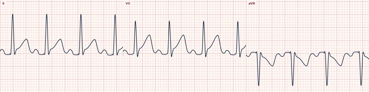
What to expect
Sinus tachycardia out of proportion to fever is frequent. Diffuse or regional ST elevation, T-wave inversion, a widening QRS, AV block and atrial or ventricular arrhythmias occur. Low voltage can appear with swelling of the heart muscle or an effusion.
Myopericarditis
When the pericardium is involved too, PR depression and diffuse ST elevation resemble pericarditis, and the troponin rises from muscle injury.
Red flags
QRS widening, high-grade AV block, VT and a falling ejection fraction mark fulminant disease and need urgent specialist care.
About the demonstration
The tracing shows sinus tachycardia with diffuse ST elevation and PR depression, as in myopericarditis.
How to read it
- Set it against the story: a recent viral illness, chest pain, breathlessness.
- Look for features of occlusion before settling on inflammation.
- Repeat the ECG for evolving conduction disease.
Pitfalls
- Focal ST elevation can be indistinguishable from infarction; coronary assessment may be needed.
- A normal ECG does not exclude myocarditis.
Frequently asked questions
Myocarditis: what is the key idea?
Myocarditis has no single ECG signature. Sinus tachycardia, nonspecific ST and T changes, ST elevation that can mimic pericarditis or infarction, conduction delay and arrhythmias are all common.
Myocarditis: what are the key numbers?
Common: Sinus tachycardia, ST and T changes; Mimics: Pericarditis, occlusion MI; Worrying signs: Wide QRS, AV block, VT.
Myocarditis: how do you apply it to an ECG?
Set it against the story: a recent viral illness, chest pain, breathlessness. Look for features of occlusion before settling on inflammation. Repeat the ECG for evolving conduction disease.
Myocarditis: what are the common pitfalls?
Focal ST elevation can be indistinguishable from infarction; coronary assessment may be needed. A normal ECG does not exclude myocarditis.