
Heart muscle and systemic disease
Draft — pending owner review
Dilated cardiomyopathy
A big, weak heart on the ECG
No single ECG sign defines dilated cardiomyopathy, but LBBB or a wide QRS, left atrial enlargement, atrial fibrillation, poor R-wave progression and ventricular ectopy together, in a breathless patient, point to it.
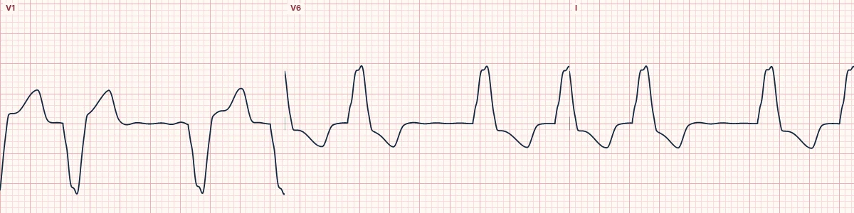
Common findings
Sinus tachycardia or atrial fibrillation, left atrial enlargement, LBBB or nonspecific QRS widening, poor R-wave progression or Q waves without an infarct, large chest-lead voltage with small limb-lead voltage, and frequent PVCs or runs of nonsustained VT.
Why they appear
Stretched, scarred muscle conducts slowly and unevenly, the enlarged left atrium fibrillates, and the scar provides circuits for ventricular arrhythmia.
Why it matters
In heart failure, LBBB with a QRS of 150 ms or more identifies patients likely to benefit from cardiac resynchronization. Nonsustained VT and syncope raise the question of a defibrillator.
About the demonstration
The tracing shows atrial fibrillation with LBBB, a common combination in advanced dilated cardiomyopathy.
How to read it
- Look for LBBB, QRS width and atrial fibrillation.
- Compare chest-lead and limb-lead voltage.
- Note ectopy and runs of VT on the rhythm strip.
Pitfalls
- LBBB makes ischemia hard to judge.
- A near-normal ECG is uncommon but does not exclude the disease.
Frequently asked questions
Dilated cardiomyopathy: what is the key idea?
No single ECG sign defines dilated cardiomyopathy, but LBBB or a wide QRS, left atrial enlargement, atrial fibrillation, poor R-wave progression and ventricular ectopy together, in a breathless patient, point to it.
Dilated cardiomyopathy: what are the key numbers?
Common: AF, LBBB, left atrial enlargement, PVCs; LBBB with QRS ≥ 150 ms: Consider resynchronization therapy; Voltage: Chest leads large relative to limb leads.
Dilated cardiomyopathy: how do you apply it to an ECG?
Look for LBBB, QRS width and atrial fibrillation. Compare chest-lead and limb-lead voltage. Note ectopy and runs of VT on the rhythm strip.
Dilated cardiomyopathy: what are the common pitfalls?
LBBB makes ischemia hard to judge. A near-normal ECG is uncommon but does not exclude the disease.