
Occlusion MI and ischemia signs
Draft — pending owner review
Pericarditis or early repolarization?
Four features that separate them
Diffuse concave ST elevation in a young person can be acute pericarditis or a normal variant. PR depression, the ST/T ratio in V6, change over time and the clinical story settle most cases.
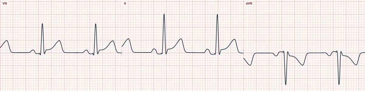
The PR segment
Pericarditis often depresses the PR segment in most leads and elevates it in aVR, from atrial inflammation. Early repolarization leaves the PR segment flat.
The ST/T ratio in V6
In V6, measure the ST elevation at the start of the ST segment and the height of the T wave, both against the PR baseline. A ratio of 0.25 or more favors pericarditis; below 0.25 favors early repolarization, whose tall T waves dwarf the ST elevation.
Distribution and change
Pericarditis tends to involve most leads, limb leads included, and evolves over days: ST elevation, then flattening, then T-wave inversion. Early repolarization is most marked in V2–V5, often has a notched J point and looks the same on every tracing.
Never forget occlusion
Convex or straight ST segments, reciprocal ST depression outside aVR and V1, new Q waves or more elevation in III than in II point to occlusion MI instead. The tracing above shows pericarditis; open Benign early repolarization, or compare the two side by side, to see the other.
How to read it
- Measure PR deviation in II and aVR.
- Work out the ST/T ratio in V6.
- Compare with a previous ECG if there is one.
Pitfalls
- The two can coexist.
- Myocarditis may accompany pericarditis and change the picture.
Frequently asked questions
Pericarditis or early repolarization?: what is the key idea?
Diffuse concave ST elevation in a young person can be acute pericarditis or a normal variant. PR depression, the ST/T ratio in V6, change over time and the clinical story settle most cases.
Pericarditis or early repolarization?: what are the key numbers?
ST/T ratio V6 ≥ 0.25: Pericarditis; ST/T ratio V6 < 0.25: Early repolarization; aVR: PR elevation in pericarditis.
Pericarditis or early repolarization?: how do you apply it to an ECG?
Measure PR deviation in II and aVR. Work out the ST/T ratio in V6. Compare with a previous ECG if there is one.
Pericarditis or early repolarization?: what are the common pitfalls?
The two can coexist. Myocarditis may accompany pericarditis and change the picture.