
Sinus
Sinus tachycardia
Sinus rhythm faster than 100/min. It is the heart's normal response to a demand such as pain, fever, blood loss or hypoxia, not a primary rhythm problem.
Last updated
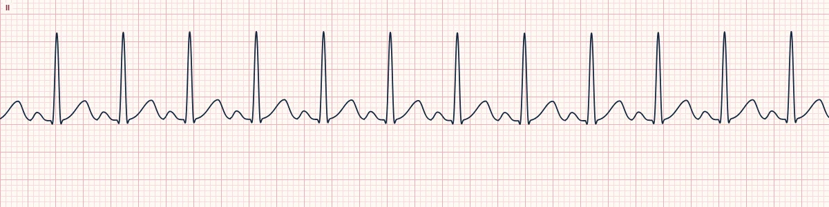
Example tracing, drawn by the lab's engine. Open the lab for a live, adjustable version.
At a glance
- Rate
- > 100/min, rarely above about 220 minus age
- Rhythm
- Regular; speeds up and slows down gradually
- P waves
- Upright in II, one before every QRS
- PR interval
- 120–200 ms, often at the short end
Figures
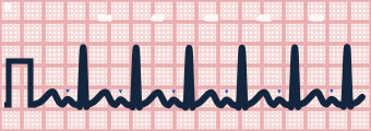
Still images drawn by the lab's engine at 25 mm/s and 10 mm/mV, with the finding labeled. Select a figure to enlarge it.
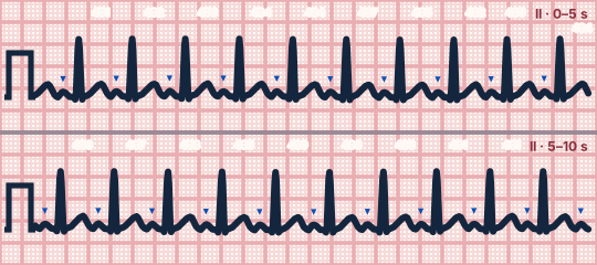
Scroll sideways to see the whole tracing, or select it to enlarge.
What the heart is doing
Sympathetic drive makes the sinus node fire faster than 100/min. Conduction through the atria, AV node and ventricles is otherwise normal, with a shorter cycle and shorter QT.
Why it looks this way
This is sinus tachycardia because upright P waves precede every narrow QRS with a constant PR, and the regular rate is above 100/min.
Why it matters
A sign, not a diagnosis. Treat the underlying cause; slowing a compensatory tachycardia with drugs or cardioversion can be harmful. An unexplained persistent rate deserves a search for bleeding, sepsis or embolism.
What to look for
- Find the P wave: at fast rates it climbs onto the end of the preceding T wave.
- Watch the rate over time. Sinus tachycardia drifts gradually; re-entrant SVT starts and stops abruptly.
- Ask what the patient needs the fast rate for before you blame the heart.
Criteria
- Rate
- > 100/min, rarely above about 220 minus age
- Rhythm
- Regular; speeds up and slows down gradually
- P waves
- Upright in II, one before every QRS
- PR interval
- 120–200 ms, often at the short end
- QRS
- Narrow (< 120 ms)
Causes
- Pain, anxiety, exertion
- Fever and sepsis
- Hypovolemia and hemorrhage
- Hypoxia, pulmonary embolism
- Stimulants, caffeine, withdrawal states
- Anemia, hyperthyroidism
Can be mistaken for
- Atrial flutter 2:1 — fixed rate near 150/min with a second flutter wave in the ST segment Compare
- AVNRT — abrupt onset, fixed rate, no visible P wave before the QRS Compare
- Focal atrial tachycardia — abnormal P-wave shape or axis Compare
Advanced Down to the ion channel
What the cells are doing
Pacemaker, atrial and ventricular action potentials, in step with the tracing above, with sodium, calcium and potassium moving through their channels.
Frequently asked questions
What are the ECG criteria for sinus tachycardia?
Rate: > 100/min, rarely above about 220 minus age. Rhythm: regular; speeds up and slows down gradually. P waves: upright in II, one before every QRS. PR interval: 120–200 ms, often at the short end. QRS: narrow (< 120 ms).
How is sinus tachycardia identified on an ECG?
This is sinus tachycardia because upright P waves precede every narrow QRS with a constant PR, and the regular rate is above 100/min.
What can sinus tachycardia be mistaken for?
Atrial flutter 2:1: fixed rate near 150/min with a second flutter wave in the ST segment. AVNRT: abrupt onset, fixed rate, no visible P wave before the QRS. Focal atrial tachycardia: abnormal P-wave shape or axis.
What causes sinus tachycardia?
Common causes: pain, anxiety, exertion; fever and sepsis; hypovolemia and hemorrhage; hypoxia, pulmonary embolism; stimulants, caffeine, withdrawal states; and anemia, hyperthyroidism.
Practise it in the ACLS Lab
Sources
This page does not list page-specific sources yet.
Every source the site cites, with links