
Waves, voltage and chambers
Draft — pending owner review
Right ventricular strain pattern
T inversion in V1–V4 and III
T-wave inversion, with or without ST depression, in the right chest leads V1–V3 (sometimes to V4) and in the inferior leads, especially III, from acute or chronic pressure overload of the right ventricle.
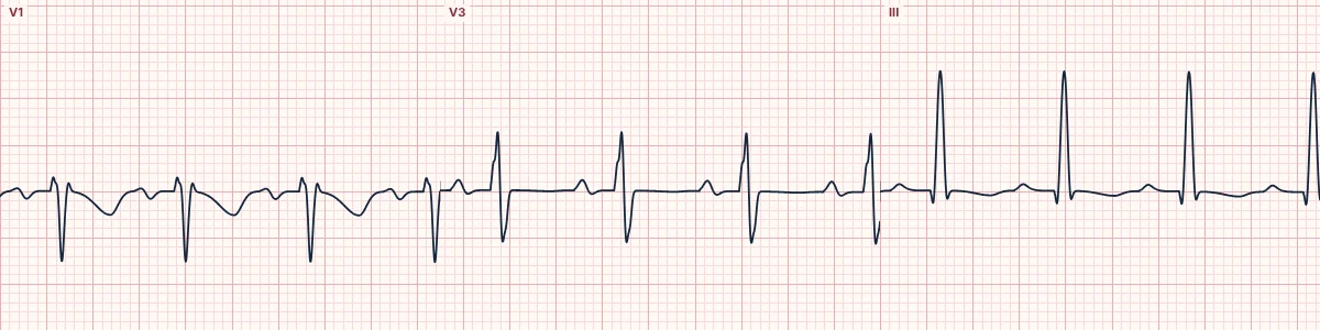
What it looks like
Inverted T waves in V1–V3, sometimes V4, and in III and aVF, often with mild ST depression. Acute strain comes with sinus tachycardia and sometimes a new RBBB, a right axis shift or an S1Q3T3 pattern.
Acute causes
Pulmonary embolism is the important one: inferior and anterior T inversion together in a breathless, tachycardic patient should raise it. Severe acute asthma or COPD and tension pneumothorax can do the same.
Chronic causes
Pulmonary hypertension, chronic thromboembolic disease and pulmonary stenosis, usually with right ventricular hypertrophy: a dominant R in V1, right axis deviation and right atrial enlargement.
Why it matters
In pulmonary embolism, T inversion in V1–V4 is linked to right ventricular dysfunction and a worse outcome. Inferior plus anterior inversion helps separate it from an anterior coronary syndrome.
How to read it
- Check whether T inversion runs across both V1–V4 and the inferior leads.
- Look for tachycardia and right-sided features.
- Put it with the patient: breathlessness, low oxygen, a swollen leg.
Pitfalls
- Wellens and anterior ischemia also invert V1–V4; inferior involvement favors the right ventricle.
- A normal ECG does not exclude pulmonary embolism.
Frequently asked questions
Right ventricular strain pattern: what is the key idea?
T-wave inversion, with or without ST depression, in the right chest leads V1–V3 (sometimes to V4) and in the inferior leads, especially III, from acute or chronic pressure overload of the right ventricle.
Right ventricular strain pattern: what are the key numbers?
Leads: V1–V3 (to V4) and III, aVF; Acute companions: Tachycardia, new RBBB, right axis shift, S1Q3T3; Chronic companions: Dominant R in V1, right axis deviation, tall P waves.
Right ventricular strain pattern: how do you apply it to an ECG?
Check whether T inversion runs across both V1–V4 and the inferior leads. Look for tachycardia and right-sided features. Put it with the patient: breathlessness, low oxygen, a swollen leg.
Right ventricular strain pattern: what are the common pitfalls?
Wellens and anterior ischemia also invert V1–V4; inferior involvement favors the right ventricle. A normal ECG does not exclude pulmonary embolism.