
Approach to tachycardia
Narrow-complex tachycardia
Regular or irregular, and where are the P waves?
A narrow QRS means the ventricles are being activated through the normal conduction system, so the rhythm starts above the ventricles. Regularity and the position of the P waves narrow the diagnosis.
Last updated
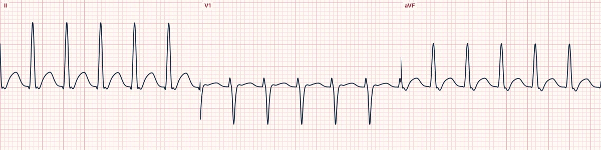
Example tracing, drawn by the lab's engine. Open the lab for a live, adjustable version.
At a glance
- Irregular
- AF, MAT, flutter with variable block
- Regular, P before QRS
- Sinus or atrial tachycardia
- Regular, rate ~150
- Consider 2:1 flutter
- No visible P
- AVNRT likely
Figures
Still images drawn by the lab's engine at 25 mm/s and 10 mm/mV, with the finding labeled. Select a figure to enlarge it.
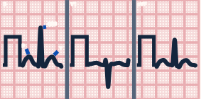
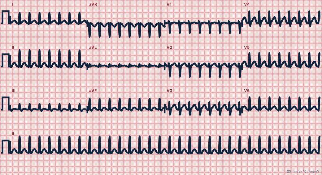
Scroll sideways to see the whole tracing, or select it to enlarge.
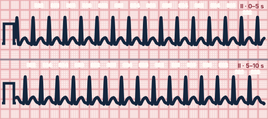
Scroll sideways to see the whole tracing, or select it to enlarge.
Irregular
Irregularly irregular with no organised atrial activity is atrial fibrillation. Irregular with at least three different P-wave shapes is multifocal atrial tachycardia. Atrial flutter with varying block is irregular but shows regular flutter waves.
Regular with visible P waves
Sinus tachycardia has normal P waves before each QRS and usually starts and stops gradually. Atrial tachycardia has abnormal P waves, often with a long RP. A rate close to 150 should always raise atrial flutter with 2:1 block.
Regular without visible P waves
AV nodal re-entry (AVNRT) hides the P wave inside or just after the QRS, sometimes as a pseudo-r′ in V1 or pseudo-S in the inferior leads. Orthodromic AV re-entry (AVRT) puts a retrograde P wave in the ST segment, more than 90 ms after the start of the QRS.
Vagal manoeuvres and adenosine
Re-entry through the AV node (AVNRT and AVRT) usually terminates. Atrial flutter and atrial tachycardia slow transiently, which unmasks the atrial activity.
How to read it
- Decide regular or irregular.
- Search every lead for P waves and note their position relative to the QRS.
- Record the rhythm during any vagal manoeuvre or adenosine.
Pitfalls
- Sinus tachycardia is usually a response to illness; treat the cause, not the rate.
Key numbers
- Irregular
- AF, MAT, flutter with variable block
- Regular, P before QRS
- Sinus or atrial tachycardia
- Regular, rate ~150
- Consider 2:1 flutter
- No visible P
- AVNRT likely
- Retrograde P in ST
- AVRT likely
Advanced Down to the ion channel
What the cells are doing
Pacemaker, atrial and ventricular action potentials, in step with the tracing above, with sodium, calcium and potassium moving through their channels.
Frequently asked questions
Narrow-complex tachycardia: what is the key idea?
A narrow QRS means the ventricles are being activated through the normal conduction system, so the rhythm starts above the ventricles. Regularity and the position of the P waves narrow the diagnosis.
Narrow-complex tachycardia: what are the key numbers?
Irregular: AF, MAT, flutter with variable block; Regular, P before QRS: Sinus or atrial tachycardia; Regular, rate ~150: Consider 2:1 flutter; No visible P: AVNRT likely; Retrograde P in ST: AVRT likely.
Narrow-complex tachycardia: how do you apply it to an ECG?
Decide regular or irregular. Search every lead for P waves and note their position relative to the QRS. Record the rhythm during any vagal manoeuvre or adenosine.
Narrow-complex tachycardia: what are the common pitfalls?
Sinus tachycardia is usually a response to illness; treat the cause, not the rate.
Sources
This page does not list page-specific sources yet.
Every source the site cites, with links