
Sinus
Sinoatrial exit block
The sinus node fires on time but the impulse fails to leave it, so a whole beat drops out. The sinus clock keeps ticking, and the next beat arrives on schedule.
Last updated
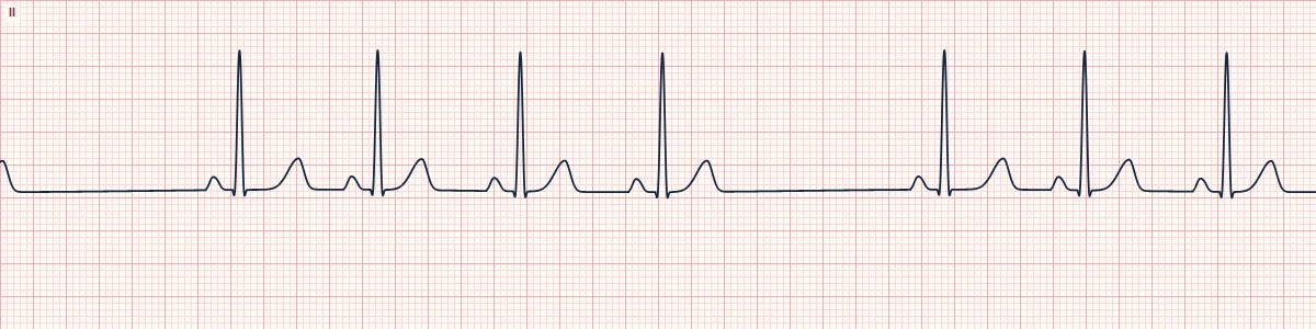
Example tracing, drawn by the lab's engine. Open the lab for a live, adjustable version.
At a glance
- Rate
- Normal or slow, with dropped beats
- Rhythm
- Regular except for the pause
- P waves
- Normal sinus P waves; one missing in the pause
- Pause
- Exactly 2 × (or 3 ×) the normal P–P interval
Figures
Still images drawn by the lab's engine at 25 mm/s and 10 mm/mV, with the finding labeled. Select a figure to enlarge it.
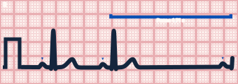
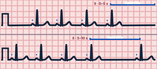
Scroll sideways to see the whole tracing, or select it to enlarge.
What the heart is doing
The sinus node depolarizes normally but the impulse is blocked in the tissue around it and never reaches the atria. Because the node's timing is undisturbed, the pause is a multiple of the sinus cycle.
Why it looks this way
This is sinoatrial exit block because a whole P–QRS–T is missing and the pause is exactly twice the normal P–P interval.
Why it matters
Often asymptomatic. Frequent or long pauses causing dizziness or syncope suggest sinus node disease; review rate-slowing drugs, and symptomatic patients may need pacing.
What to look for
- March the P waves through the pause: the next P lands exactly where the beat after the missing one should be.
- An entire P–QRS–T is missing, unlike AV block where the P wave is present.
- The PR of the beats around the pause is unchanged.
Criteria
- Rate
- Normal or slow, with dropped beats
- Rhythm
- Regular except for the pause
- P waves
- Normal sinus P waves; one missing in the pause
- Pause
- Exactly 2 × (or 3 ×) the normal P–P interval
- QRS
- Narrow (< 120 ms)
Causes
- Sinus node dysfunction (sick sinus syndrome)
- High vagal tone
- Digoxin, beta-blockers, calcium-channel blockers
- Inferior myocardial infarction
- Myocarditis
- Hyperkalemia
Can be mistaken for
- Sinus pause — the gap is not a multiple of the P–P interval Compare
- Non-conducted PAC — an early, differently shaped P wave deforms the T wave
- Sinus arrhythmia — the interval changes gradually, not by a whole beat Compare
Advanced Down to the ion channel
What the cells are doing
Pacemaker, atrial and ventricular action potentials, in step with the tracing above, with sodium, calcium and potassium moving through their channels.
Frequently asked questions
What are the ECG criteria for sinoatrial exit block?
Rate: normal or slow, with dropped beats. Rhythm: regular except for the pause. P waves: normal sinus P waves; one missing in the pause. Pause: exactly 2 × (or 3 ×) the normal P–P interval. QRS: narrow (< 120 ms).
How is sinoatrial exit block identified on an ECG?
This is sinoatrial exit block because a whole P–QRS–T is missing and the pause is exactly twice the normal P–P interval.
What can sinoatrial exit block be mistaken for?
Sinus pause: the gap is not a multiple of the P–P interval. Non-conducted PAC: an early, differently shaped P wave deforms the T wave. Sinus arrhythmia: the interval changes gradually, not by a whole beat.
What causes sinoatrial exit block?
Common causes: sinus node dysfunction (sick sinus syndrome); high vagal tone; digoxin, beta-blockers, calcium-channel blockers; inferior myocardial infarction; myocarditis; and hyperkalemia.
Sources
This page does not list page-specific sources yet.
Every source the site cites, with links