
Sinus
Normal sinus rhythm
The heart's normal rhythm: every beat starts in the sinus node and travels the usual pathway to the ventricles at 60–100/min.
Last updated
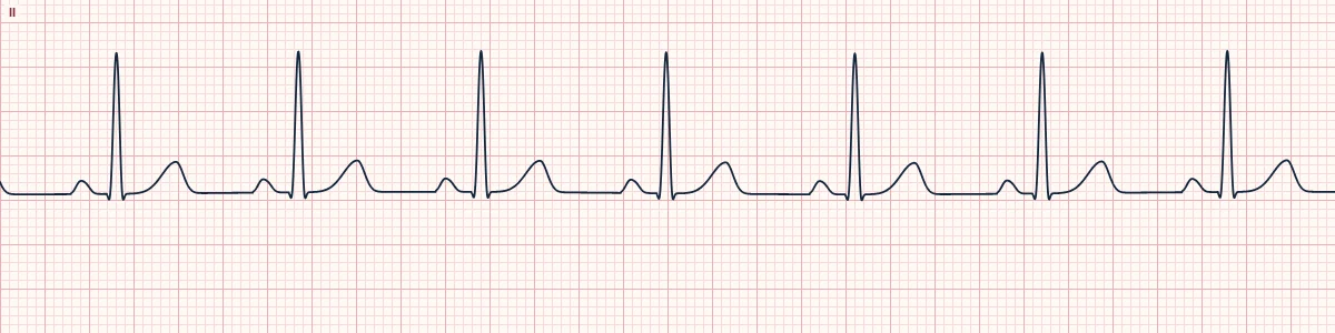
Example tracing, drawn by the lab's engine. Open the lab for a live, adjustable version.
At a glance
- Rate
- 60–100/min
- Rhythm
- Regular
- P waves
- Upright in II, one before every QRS
- PR interval
- 120–200 ms, constant
Figures
Still images drawn by the lab's engine at 25 mm/s and 10 mm/mV, with the finding labeled. Select a figure to enlarge it.
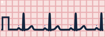
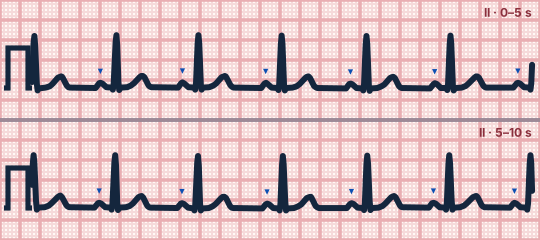
Scroll sideways to see the whole tracing, or select it to enlarge.
What the heart is doing
The sinus node fires 60–100 times a minute. The impulse crosses both atria, is briefly delayed in the AV node, then spreads rapidly through the His–Purkinje system to both ventricles.
Why it looks this way
This is normal sinus rhythm because the rate is 60–100/min and regular, with an upright P wave and a constant normal PR before every narrow QRS.
Why it matters
The reference rhythm that every other strip is compared with. It names the rhythm only: it does not mean the rest of the ECG is normal, so still read the axis, intervals, QRS, ST and T. Nor does it exclude shock or a sick patient.
What to look for
- Check that every QRS has a P wave in front of it and every P wave is followed by a QRS.
- P waves should be upright in I and II and inverted in aVR; if not, suspect a non-sinus focus or misplaced limb leads.
- March out the R–R intervals with calipers or paper: they should be essentially equal.
Criteria
- Rate
- 60–100/min
- Rhythm
- Regular
- P waves
- Upright in II, one before every QRS
- PR interval
- 120–200 ms, constant
- QRS
- Narrow (< 120 ms)
Causes
- Normal resting state
- Healthy sinus node and conduction system
- Rhythm restored after a treated arrhythmia
Can be mistaken for
- Ectopic atrial rhythm — P waves of a different shape or axis from sinus; inverted in II only when the focus is low Compare
- Atrial flutter 4:1 — sawtooth baseline instead of a flat line between beats Compare
- Accelerated junctional rhythm — retrograde P waves before the QRS (PR < 120 ms), hidden within it or after it Compare
Advanced Down to the ion channel
What the cells are doing
Pacemaker, atrial and ventricular action potentials, in step with the tracing above, with sodium, calcium and potassium moving through their channels.
Frequently asked questions
What are the ECG criteria for normal sinus rhythm?
Rate: 60–100/min. Rhythm: regular. P waves: upright in II, one before every QRS. PR interval: 120–200 ms, constant. QRS: narrow (< 120 ms).
How is normal sinus rhythm identified on an ECG?
This is normal sinus rhythm because the rate is 60–100/min and regular, with an upright P wave and a constant normal PR before every narrow QRS.
What can normal sinus rhythm be mistaken for?
Ectopic atrial rhythm: P waves of a different shape or axis from sinus; inverted in II only when the focus is low. Atrial flutter 4:1: sawtooth baseline instead of a flat line between beats. Accelerated junctional rhythm: retrograde P waves before the QRS (PR < 120 ms), hidden within it or after it.
What causes normal sinus rhythm?
Common causes: normal resting state; healthy sinus node and conduction system; and rhythm restored after a treated arrhythmia.
Practise it in the ACLS Lab
Sources
This page does not list page-specific sources yet.
Every source the site cites, with links