
Sinus
Sinus arrhythmia
Sinus rhythm whose rate rises and falls in a cycle, usually with breathing. A normal finding, most obvious in children and fit young adults.
Last updated
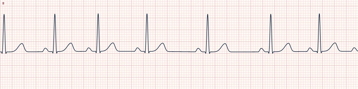
Example tracing, drawn by the lab's engine. Open the lab for a live, adjustable version.
At a glance
- Rate
- Usually 60–100/min, varying cyclically
- Rhythm
- Regularly irregular; P–P varies by > 120 ms
- P waves
- Upright in II, identical in shape
- PR interval
- 120–200 ms, constant
Figures
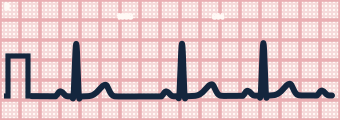
Still images drawn by the lab's engine at 25 mm/s and 10 mm/mV, with the finding labeled. Select a figure to enlarge it.
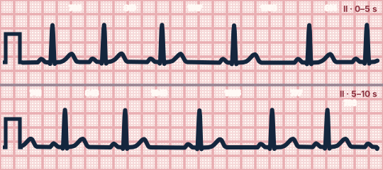
Scroll sideways to see the whole tracing, or select it to enlarge.
What the heart is doing
Breathing changes vagal tone on the sinus node: inspiration reduces it and the node fires faster, expiration restores it and the node slows. Conduction below the node is unchanged.
Why it looks this way
This is sinus arrhythmia because identical sinus P waves with a constant PR precede every QRS while the P–P interval waxes and wanes cyclically.
Why it matters
Benign and needs no treatment. Its main importance is being told apart from pathological irregular rhythms such as atrial fibrillation or sinus node disease.
What to look for
- The R–R intervals shorten and lengthen smoothly over several beats rather than jumping suddenly.
- Every P wave looks the same and has the same PR; only the spacing changes.
- The rate speeds up on inspiration and slows on expiration.
Criteria
- Rate
- Usually 60–100/min, varying cyclically
- Rhythm
- Regularly irregular; P–P varies by > 120 ms
- P waves
- Upright in II, identical in shape
- PR interval
- 120–200 ms, constant
- QRS
- Narrow (< 120 ms)
Causes
- Normal respiratory variation in vagal tone
- Youth and athletic conditioning
- Sleep
- Recovery after exercise
- Digoxin or opioid effect (non-respiratory form)
Can be mistaken for
- Wandering atrial pacemaker — the P-wave shape and PR change from beat to beat Compare
- Premature atrial complexes — a single early beat with a different P wave Compare
- Sinoatrial exit block — a sudden pause exactly two cycles long Compare
Advanced Down to the ion channel
What the cells are doing
Pacemaker, atrial and ventricular action potentials, in step with the tracing above, with sodium, calcium and potassium moving through their channels.
Frequently asked questions
What are the ECG criteria for sinus arrhythmia?
Rate: usually 60–100/min, varying cyclically. Rhythm: regularly irregular; P–P varies by > 120 ms. P waves: upright in II, identical in shape. PR interval: 120–200 ms, constant. QRS: narrow (< 120 ms).
How is sinus arrhythmia identified on an ECG?
This is sinus arrhythmia because identical sinus P waves with a constant PR precede every QRS while the P–P interval waxes and wanes cyclically.
What can sinus arrhythmia be mistaken for?
Wandering atrial pacemaker: the P-wave shape and PR change from beat to beat. Premature atrial complexes: a single early beat with a different P wave. Sinoatrial exit block: a sudden pause exactly two cycles long.
What causes sinus arrhythmia?
Common causes: normal respiratory variation in vagal tone; youth and athletic conditioning; sleep; recovery after exercise; and digoxin or opioid effect (non-respiratory form).
Sources
This page does not list page-specific sources yet.
Every source the site cites, with links