
Ventricular
Draft — pending owner review
Ventricular flutter
A very fast ventricular rhythm, about 250–350/min, in which the QRS and T wave merge into a regular sine-like wave with no flat baseline. It is usually pulseless and often becomes VF.
Last updated
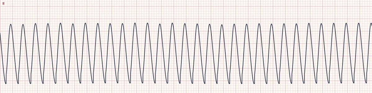
Example tracing, drawn by the lab's engine. Open the lab for a live, adjustable version.
At a glance
- Rate
- About 250–350/min
- Waveform
- Regular, uniform sine-like oscillations
- Baseline
- No isoelectric line; QRS and T cannot be separated
- P waves
- Not seen
Figures
Still images drawn by the lab's engine at 25 mm/s and 10 mm/mV, with the finding labeled. Select a figure to enlarge it.
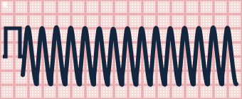
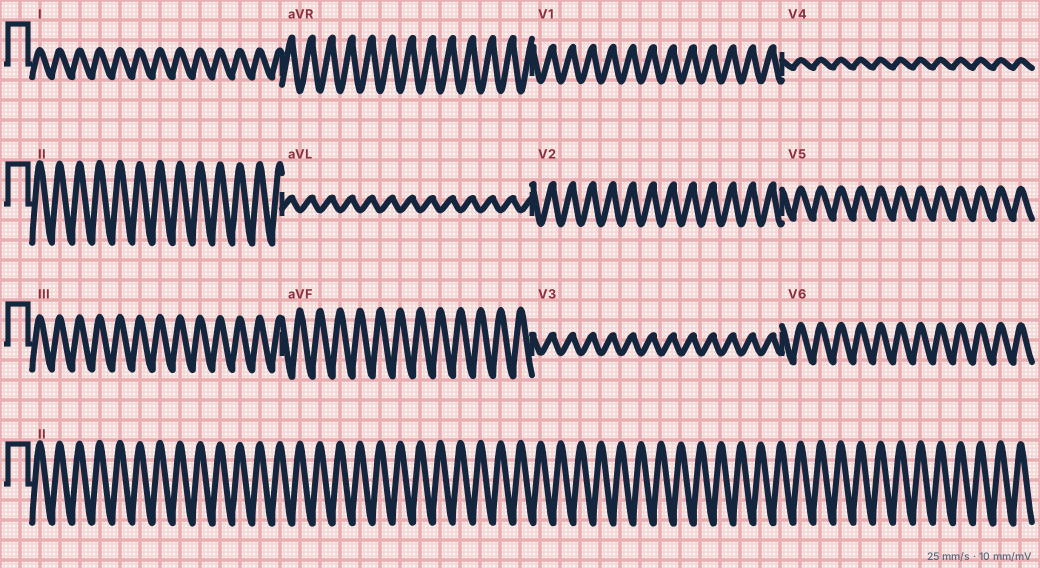
Scroll sideways to see the whole tracing, or select it to enlarge.
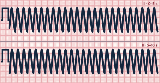
Scroll sideways to see the whole tracing, or select it to enlarge.
What the heart is doing
A single very rapid re-entrant circuit activates the ventricles so often that repolarization never finishes before the next depolarization begins, so the waves run into each other.
Why it looks this way
This is ventricular flutter because regular, uniform sine-like waves at about 300/min have no isoelectric baseline and no separable QRS or T wave.
Why it matters
Without a pulse it is a cardiac arrest rhythm, treated like VF and pulseless VT. It is unstable and usually degenerates into VF within seconds to minutes.
What to look for
- Large regular waves with no flat segment anywhere.
- You cannot say which part is the QRS and which the T wave.
- Check the patient: a pulse is usually absent.
Criteria
- Rate
- About 250–350/min
- Waveform
- Regular, uniform sine-like oscillations
- Baseline
- No isoelectric line; QRS and T cannot be separated
- P waves
- Not seen
- Patient
- Usually no pulse
Causes
- Acute ischemia or infarction
- Scar-related VT accelerating
- Severe electrolyte disturbance
- Drug toxicity
- Electrical injury
Can be mistaken for
- Monomorphic VT — slower, with distinct QRS complexes and T waves Compare
- Coarse VF — irregular in size, shape and timing Compare
- Severe hyperkalemia — sine-wave pattern at a slower rate Compare
Advanced Down to the ion channel
What the cells are doing
Pacemaker, atrial and ventricular action potentials, in step with the tracing above, with sodium, calcium and potassium moving through their channels.
Frequently asked questions
What are the ECG criteria for ventricular flutter?
Rate: about 250–350/min. Waveform: regular, uniform sine-like oscillations. Baseline: no isoelectric line; QRS and T cannot be separated. P waves: not seen. Patient: usually no pulse.
How is ventricular flutter identified on an ECG?
This is ventricular flutter because regular, uniform sine-like waves at about 300/min have no isoelectric baseline and no separable QRS or T wave.
What can ventricular flutter be mistaken for?
Monomorphic VT: slower, with distinct QRS complexes and T waves. Coarse VF: irregular in size, shape and timing. Severe hyperkalemia: sine-wave pattern at a slower rate.
What causes ventricular flutter?
Common causes: acute ischemia or infarction; scar-related VT accelerating; severe electrolyte disturbance; drug toxicity; and electrical injury.
Sources
This page does not list page-specific sources yet.
Every source the site cites, with links