
Ventricular
Torsades de pointes
A form of polymorphic VT linked to a long QT interval. The complexes swell and shrink as if twisting around the baseline.
Last updated
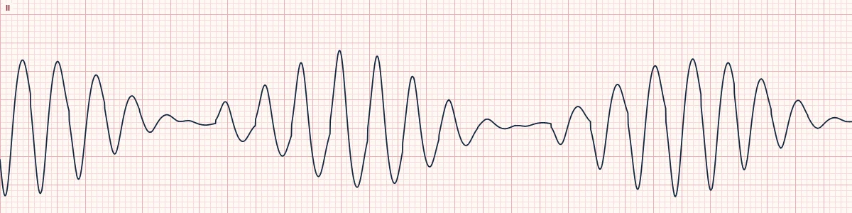
Example tracing, drawn by the lab's engine. Open the lab for a live, adjustable version.
At a glance
- Rate
- Usually 200–250/min
- Rhythm
- Irregular
- QRS
- Wide; amplitude waxes and wanes in spindles
- Axis
- Rotates around the baseline every 5–20 beats
Figures
Still images drawn by the lab's engine at 25 mm/s and 10 mm/mV, with the finding labeled. Select a figure to enlarge it.
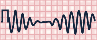
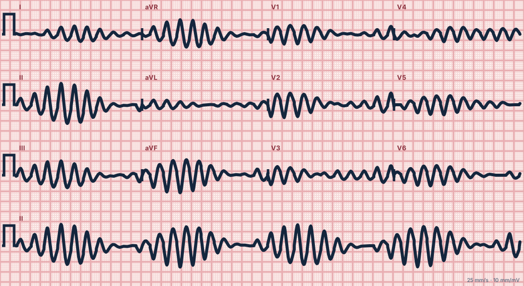
Scroll sideways to see the whole tracing, or select it to enlarge.
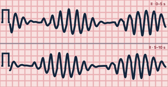
Scroll sideways to see the whole tracing, or select it to enlarge.
What the heart is doing
Prolonged repolarization allows early afterdepolarizations that trigger a beat during the T wave. Uneven recovery across the ventricle then sustains a drifting re-entrant wave, which rotates the QRS axis.
Why it looks this way
This is torsades de pointes because the wide complexes twist around the baseline in spindles, the hallmark of polymorphic VT with a long QT.
Why it matters
Often comes in self-terminating bursts but can become VF. A sustained episode needs an immediate unsynchronized shock, with or without a pulse. Bursts that stop on their own are not shocked: give magnesium, stop QT-prolonging drugs, correct electrolytes and prevent slow rates.
What to look for
- A spindle or ribbon shape: complexes grow, shrink to almost nothing, then grow in the opposite direction.
- It often starts after a pause, with a PVC landing on a long, late T wave.
- Measure the QT on sinus beats between runs.
Criteria
- Rate
- Usually 200–250/min
- Rhythm
- Irregular
- QRS
- Wide; amplitude waxes and wanes in spindles
- Axis
- Rotates around the baseline every 5–20 beats
- Baseline QT
- Prolonged (QTc usually > 500 ms)
Causes
- QT-prolonging drugs (antiarrhythmics, antipsychotics, some antibiotics)
- Hypokalemia
- Hypomagnesemia
- Hypocalcemia
- Congenital long QT syndrome
- Marked bradycardia or AV block
Can be mistaken for
- Polymorphic VT with normal QT — chaotic changes without a smooth twist; think ischemia Compare
- Coarse ventricular fibrillation — no organized complexes or spindles Compare
- Artifact from movement — underlying QRS complexes march through
Advanced Down to the ion channel
What the cells are doing
Pacemaker, atrial and ventricular action potentials, in step with the tracing above, with sodium, calcium and potassium moving through their channels.
Frequently asked questions
What are the ECG criteria for torsades de pointes?
Rate: usually 200–250/min. Rhythm: irregular. QRS: wide; amplitude waxes and wanes in spindles. Axis: rotates around the baseline every 5–20 beats. Baseline QT: prolonged (QTc usually > 500 ms).
How is torsades de pointes identified on an ECG?
This is torsades de pointes because the wide complexes twist around the baseline in spindles, the hallmark of polymorphic VT with a long QT.
What can torsades de pointes be mistaken for?
Polymorphic VT with normal QT: chaotic changes without a smooth twist; think ischemia. Coarse ventricular fibrillation: no organized complexes or spindles. Artifact from movement: underlying QRS complexes march through.
What causes torsades de pointes?
Common causes: QT-prolonging drugs (antiarrhythmics, antipsychotics, some antibiotics); hypokalemia; hypomagnesemia; hypocalcemia; congenital long QT syndrome; and marked bradycardia or AV block.
Practise it in the ACLS Lab
Sources
This page does not list page-specific sources yet.
Every source the site cites, with links