
Chamber enlargement
Apical hypertrophic cardiomyopathy
A form of hypertrophic cardiomyopathy in which the thickening is concentrated at the tip of the left ventricle, producing giant T-wave inversions in the chest leads.
Last updated
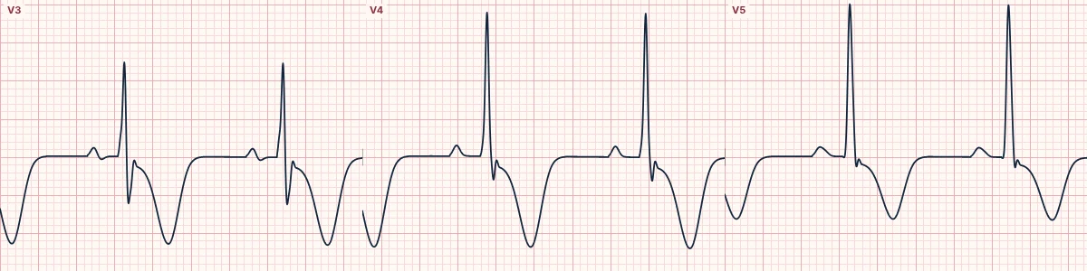
Example tracing, drawn by the lab's engine. Open the lab for a live, adjustable version.
At a glance
- T waves
- Deep, symmetric inversion (classically ≥ 10 mm) in V3–V5
- QRS
- High voltage in the precordial leads
- ST segment
- Mild depression in the same leads
- Leads
- Mid-precordial, often lateral and inferior too
Figures
Still images drawn by the lab's engine at 25 mm/s and 10 mm/mV, with the finding labeled. Select a figure to enlarge it.
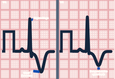
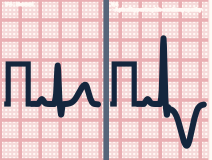
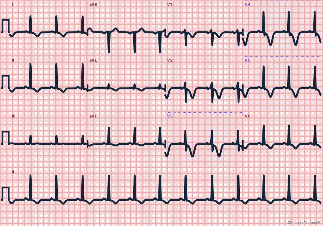
Scroll sideways to see the whole tracing, or select it to enlarge.
What the heart is doing
Thick apical muscle generates large forces toward the mid-precordial leads and repolarizes in reverse order, so the T-wave vector points away from the apex and inverts deeply.
Why it looks this way
This is apical hypertrophic cardiomyopathy because high precordial voltage is combined with giant symmetric T-wave inversions in the mid-chest leads.
Why it matters
Generally milder than other forms of hypertrophic cardiomyopathy but can cause atrial fibrillation, apical aneurysm and ventricular arrhythmias. It imitates ischemia: confirm with imaging.
What to look for
- Very tall R waves in V4–V5 sitting above very deep, symmetric T inversions.
- The pattern is stable from one ECG to the next, unlike evolving ischemia.
- No Q waves and no loss of R waves.
Criteria
- T waves
- Deep, symmetric inversion (classically ≥ 10 mm) in V3–V5
- QRS
- High voltage in the precordial leads
- ST segment
- Mild depression in the same leads
- Leads
- Mid-precordial, often lateral and inferior too
Causes
- Inherited sarcomere gene variants
- Family history of cardiomyopathy
- More prevalent in East Asian populations
- Often found incidentally in adults
Can be mistaken for
- Wellens pattern type B — normal voltage, recent chest pain, changes evolve Compare
- Raised intracranial pressure — broad T inversions with a very long QT Compare
- LVH with strain — asymmetric, shallower T inversion in lateral leads Compare
Advanced Down to the ion channel
What the cells are doing
Pacemaker, atrial and ventricular action potentials, in step with the tracing above, with sodium, calcium and potassium moving through their channels.
Frequently asked questions
What are the ECG criteria for apical hypertrophic cardiomyopathy?
T waves: deep, symmetric inversion (classically ≥ 10 mm) in V3–V5. QRS: high voltage in the precordial leads. ST segment: mild depression in the same leads. Leads: mid-precordial, often lateral and inferior too.
How is apical hypertrophic cardiomyopathy identified on an ECG?
This is apical hypertrophic cardiomyopathy because high precordial voltage is combined with giant symmetric T-wave inversions in the mid-chest leads.
What can apical hypertrophic cardiomyopathy be mistaken for?
Wellens pattern type B: normal voltage, recent chest pain, changes evolve. Raised intracranial pressure: broad T inversions with a very long QT. LVH with strain: asymmetric, shallower T inversion in lateral leads.
What causes apical hypertrophic cardiomyopathy?
Common causes: inherited sarcomere gene variants; family history of cardiomyopathy; more prevalent in East Asian populations; and often found incidentally in adults.
Sources
This page does not list page-specific sources yet.
Every source the site cites, with links