
Other conditions
Raised intracranial pressure (cerebral T waves)
Severe brain injury triggers a surge in autonomic activity that distorts repolarization, giving giant, wide, inverted T waves and a very long QT.
Last updated
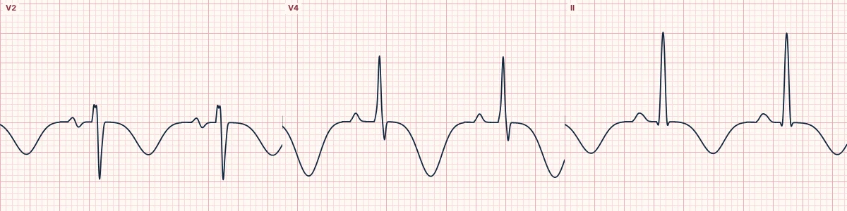
Example tracing, drawn by the lab's engine. Open the lab for a live, adjustable version.
At a glance
- T waves
- Deep, wide, symmetric inversion in most leads
- QT
- Markedly prolonged (QTc often > 500 ms)
- Leads
- Chest leads most marked; limb leads too
- Rate
- Often bradycardia
Figures
Still images drawn by the lab's engine at 25 mm/s and 10 mm/mV, with the finding labeled. Select a figure to enlarge it.
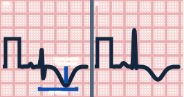
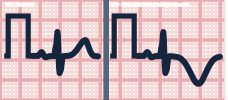
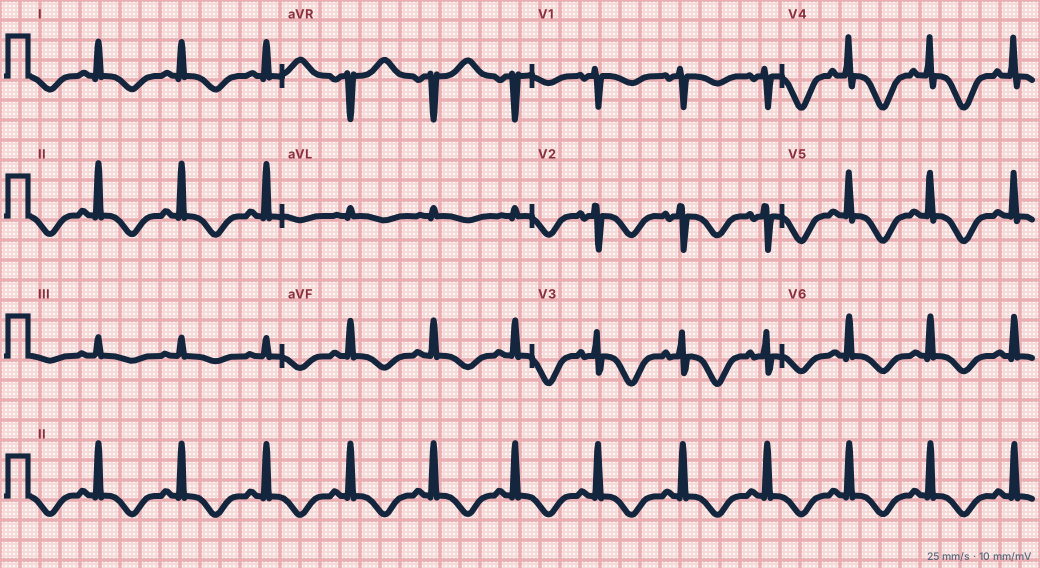
Scroll sideways to see the whole tracing, or select it to enlarge.
What the heart is doing
Intense sympathetic discharge from the injured brain alters ventricular repolarization unevenly and can stun the muscle. Repolarization is delayed and reversed, widening and inverting the T wave.
Why it looks this way
These are cerebral T waves because the T-wave inversions are deep, wide and widespread and the QT is markedly prolonged.
Why it matters
The ECG reflects the brain, not a blocked coronary artery. Prioritize neurological assessment and airway; hypertension, bradycardia and irregular breathing together warn of impending herniation.
What to look for
- T inversions that are unusually broad as well as deep.
- A very long QT accompanying the inversions.
- Headache, reduced consciousness or neurological signs in place of chest pain.
Criteria
- T waves
- Deep, wide, symmetric inversion in most leads
- QT
- Markedly prolonged (QTc often > 500 ms)
- Leads
- Chest leads most marked; limb leads too
- Rate
- Often bradycardia
Causes
- Subarachnoid hemorrhage
- Intracerebral hemorrhage
- Large ischemic stroke
- Traumatic brain injury
- Brain tumor or hydrocephalus
Can be mistaken for
- Wellens pattern type B — narrower inversions in V2–V4 with a near-normal QT Compare
- Apical hypertrophic cardiomyopathy — high voltage with a normal QT Compare
- Ischemia after tachycardia or pacing — history gives the answer
Advanced Down to the ion channel
What the cells are doing
Pacemaker, atrial and ventricular action potentials, in step with the tracing above, with sodium, calcium and potassium moving through their channels.
Frequently asked questions
What are the ECG criteria for raised intracranial pressure (cerebral T waves)?
T waves: deep, wide, symmetric inversion in most leads. QT: markedly prolonged (QTc often > 500 ms). Leads: chest leads most marked; limb leads too. Rate: often bradycardia.
How is raised intracranial pressure (cerebral T waves) identified on an ECG?
These are cerebral T waves because the T-wave inversions are deep, wide and widespread and the QT is markedly prolonged.
What can raised intracranial pressure (cerebral T waves) be mistaken for?
Wellens pattern type B: narrower inversions in V2–V4 with a near-normal QT. Apical hypertrophic cardiomyopathy: high voltage with a normal QT. Ischemia after tachycardia or pacing: history gives the answer.
What causes raised intracranial pressure (cerebral T waves)?
Common causes: subarachnoid hemorrhage; intracerebral hemorrhage; large ischemic stroke; traumatic brain injury; and brain tumor or hydrocephalus.
Sources
This page does not list page-specific sources yet.
Every source the site cites, with links