
Other conditions
Hypothermia (Osborn waves)
A cold heart conducts and repolarizes slowly. The hallmark is the Osborn (J) wave, an extra hump at the end of the QRS, along with long intervals.
Last updated
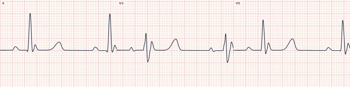
Example tracing, drawn by the lab's engine. Open the lab for a live, adjustable version.
At a glance
- J point
- Osborn wave: a positive hump at the end of the QRS
- Leads
- Best in V2–V5 and the inferior leads
- Intervals
- PR, QRS and QT all prolonged
- Rate
- Bradycardia; slow atrial fibrillation is common
Figures
Still images drawn by the lab's engine at 25 mm/s and 10 mm/mV, with the finding labeled. Select a figure to enlarge it.
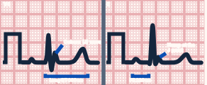
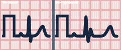
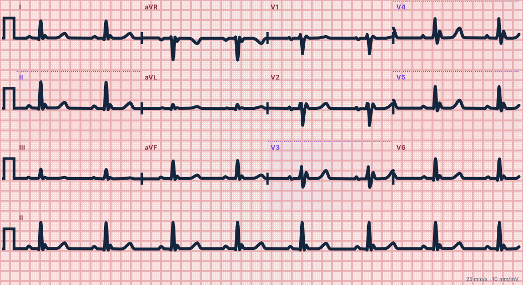
Scroll sideways to see the whole tracing, or select it to enlarge.
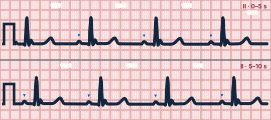
Scroll sideways to see the whole tracing, or select it to enlarge.
What the heart is doing
Cold slows all ion channels. A larger early-repolarization gradient between the outer and inner ventricular wall inscribes the J wave, and slowed conduction lengthens every interval.
Why it looks this way
This is hypothermia because Osborn waves appear at the J point together with prolonged PR, QRS and QT intervals.
Why it matters
Below about 30 °C the heart is irritable and rough handling can provoke ventricular fibrillation. Handle gently and rewarm; the bradycardia is appropriate and usually needs no pacing or drugs.
What to look for
- A dome-shaped extra wave between the QRS and the ST segment.
- The colder the patient, the larger the Osborn wave.
- A fuzzy baseline from shivering, which disappears in severe hypothermia.
Criteria
- J point
- Osborn wave: a positive hump at the end of the QRS
- Leads
- Best in V2–V5 and the inferior leads
- Intervals
- PR, QRS and QT all prolonged
- Rate
- Bradycardia; slow atrial fibrillation is common
- Baseline
- Shivering artifact
Causes
- Environmental cold exposure
- Cold-water immersion
- Intoxication or overdose with exposure
- Sepsis
- Hypothyroidism
- Frailty and trauma
Can be mistaken for
- Early repolarization — a small J notch with normal intervals Compare
- Brugada pattern — coved elevation limited to V1–V2 Compare
- STEMI — ST elevation without a discrete hump, with reciprocal changes
Advanced Down to the ion channel
What the cells are doing
Pacemaker, atrial and ventricular action potentials, in step with the tracing above, with sodium, calcium and potassium moving through their channels.
Frequently asked questions
What are the ECG criteria for hypothermia (Osborn waves)?
J point: osborn wave: a positive hump at the end of the QRS. Leads: best in V2–V5 and the inferior leads. Intervals: PR, QRS and QT all prolonged. Rate: bradycardia; slow atrial fibrillation is common. Baseline: shivering artifact.
How is hypothermia (Osborn waves) identified on an ECG?
This is hypothermia because Osborn waves appear at the J point together with prolonged PR, QRS and QT intervals.
What can hypothermia (Osborn waves) be mistaken for?
Early repolarization: a small J notch with normal intervals. Brugada pattern: coved elevation limited to V1–V2. STEMI: ST elevation without a discrete hump, with reciprocal changes.
What causes hypothermia (Osborn waves)?
Common causes: environmental cold exposure; cold-water immersion; intoxication or overdose with exposure; sepsis; hypothyroidism; and frailty and trauma.
Practise it in the ACLS Lab
Sources
This page does not list page-specific sources yet.
Every source the site cites, with links