
Other conditions
Pericardial effusion with electrical alternans
A large collection of fluid around the heart dampens the ECG signal and lets the heart swing within it, so QRS height and axis alternate from beat to beat.
Last updated
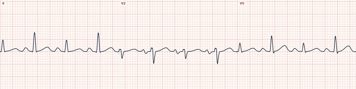
Example tracing, drawn by the lab's engine. Open the lab for a live, adjustable version.
At a glance
- QRS voltage
- Low: < 5 mm in limb leads, < 10 mm in chest leads
- Electrical alternans
- QRS height and axis alternate every other beat
- Rate
- Usually sinus tachycardia
- ST–T
- Nonspecific, or changes of pericarditis
Figures
Still images drawn by the lab's engine at 25 mm/s and 10 mm/mV, with the finding labeled. Select a figure to enlarge it.
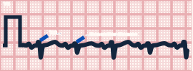
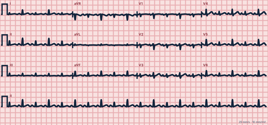
Scroll sideways to see the whole tracing, or select it to enlarge.
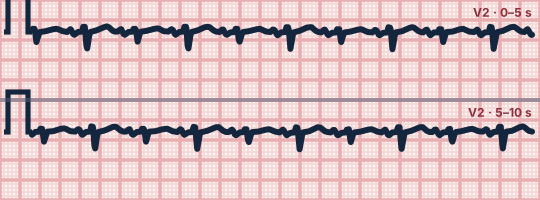
Scroll sideways to see the whole tracing, or select it to enlarge.
What the heart is doing
Fluid between the heart and chest wall shunts current and lowers the recorded voltage. The heart swings to and fro in the fluid with each beat, changing its position relative to the electrodes.
Why it looks this way
This is a pericardial effusion with electrical alternans because the QRS is low in voltage and alternates in height from beat to beat.
Why it matters
With hypotension, distended neck veins or muffled heart sounds this is cardiac tamponade, a form of obstructive shock. Urgent drainage is the treatment; fluids only buy time.
What to look for
- Compare each QRS with the next: tall–short–tall–short in a regular rhythm.
- Complexes are small in all leads.
- A fast rate with small, alternating complexes should prompt a search for tamponade.
Criteria
- QRS voltage
- Low: < 5 mm in limb leads, < 10 mm in chest leads
- Electrical alternans
- QRS height and axis alternate every other beat
- Rate
- Usually sinus tachycardia
- ST–T
- Nonspecific, or changes of pericarditis
Causes
- Cancer involving the pericardium
- Pericarditis (viral, uremic, tuberculous)
- Chest trauma
- Aortic dissection
- After cardiac surgery or procedures
- Rupture after myocardial infarction
Can be mistaken for
- Low voltage from obesity, emphysema or hypothyroidism — no alternans
- QRS alternans in rapid SVT — very fast rate with normal voltage
- Ventricular bigeminy — alternate beats are early and wide Compare
Advanced Down to the ion channel
What the cells are doing
Pacemaker, atrial and ventricular action potentials, in step with the tracing above, with sodium, calcium and potassium moving through their channels.
Frequently asked questions
What are the ECG criteria for pericardial effusion with electrical alternans?
QRS voltage: low: < 5 mm in limb leads, < 10 mm in chest leads. Electrical alternans: QRS height and axis alternate every other beat. Rate: usually sinus tachycardia. ST–T: nonspecific, or changes of pericarditis.
How is pericardial effusion with electrical alternans identified on an ECG?
This is a pericardial effusion with electrical alternans because the QRS is low in voltage and alternates in height from beat to beat.
What can pericardial effusion with electrical alternans be mistaken for?
Low voltage from obesity, emphysema or hypothyroidism: no alternans. QRS alternans in rapid SVT: very fast rate with normal voltage. Ventricular bigeminy: alternate beats are early and wide.
What causes pericardial effusion with electrical alternans?
Common causes: cancer involving the pericardium; pericarditis (viral, uremic, tuberculous); chest trauma; aortic dissection; after cardiac surgery or procedures; and rupture after myocardial infarction.
Sources
This page does not list page-specific sources yet.
Every source the site cites, with links