
Other conditions
Draft — pending owner review
Takotsubo (stress) cardiomyopathy
Stress cardiomyopathy: sudden apical ballooning, often after emotional or physical stress. The ECG mimics an anterior STEMI, then evolves to deep T inversion with a long QT.
Last updated
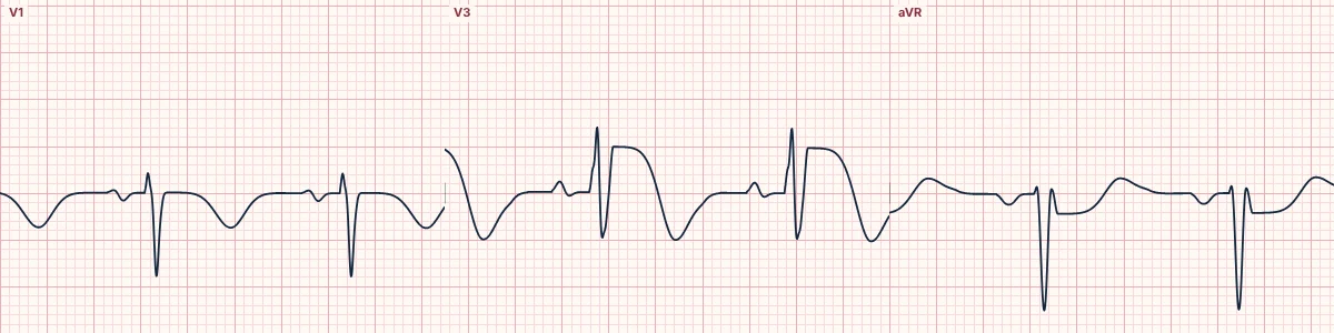
Example tracing, drawn by the lab's engine. Open the lab for a live, adjustable version.
At a glance
- ST elevation
- V2–V5, often also lateral and inferior; usually little in V1
- aVR
- ST depression is common
- Inferior leads
- Usually no reciprocal ST depression
- T waves
- Deep, broad, symmetric inversion over days
Figures
Still images drawn by the lab's engine at 25 mm/s and 10 mm/mV, with the finding labeled. Select a figure to enlarge it.
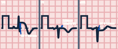
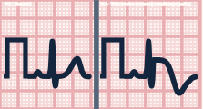
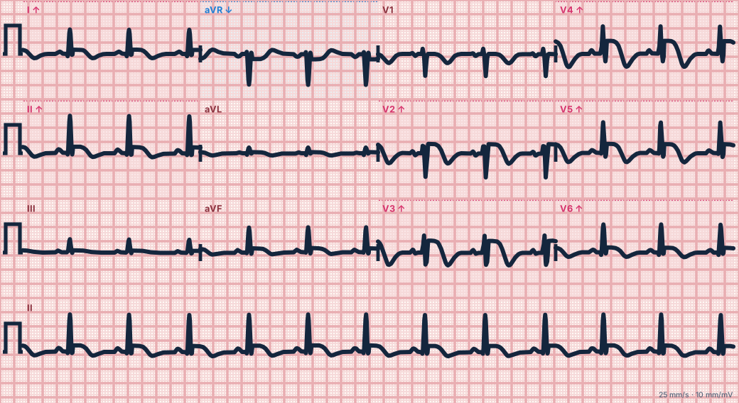
Scroll sideways to see the whole tracing, or select it to enlarge.
What the heart is doing
A catecholamine surge stuns the apical and mid left ventricle, which balloons while the base keeps contracting. The injury follows the apex rather than one coronary artery, so ST changes spread across territories.
Why it looks this way
This is Takotsubo cardiomyopathy because ST elevation spans the anterior and lateral leads without inferior reciprocal depression, with deep symmetric T inversion and a long QT.
Why it matters
Indistinguishable from an anterior STEMI on the ECG, so it needs urgent coronary angiography. Complications include shock, outflow obstruction, LV thrombus and torsades during the long-QT phase. Most patients recover.
What to look for
- Treat it as an anterior STEMI until the coronary arteries are seen: the ECG cannot exclude occlusion.
- Note widespread changes beyond one coronary territory, with ST depression in aVR.
- Measure the QTc; deep T inversion with a long QT carries a risk of torsades.
Criteria
- ST elevation
- V2–V5, often also lateral and inferior; usually little in V1
- aVR
- ST depression is common
- Inferior leads
- Usually no reciprocal ST depression
- T waves
- Deep, broad, symmetric inversion over days
- QTc
- Prolonged, often > 500 ms
Causes
- Emotional stress (bereavement, conflict)
- Physical stress (surgery, sepsis, asthma)
- Neurological events (subarachnoid hemorrhage, seizure)
- Catecholamine excess or stimulants
- Most often postmenopausal women
Can be mistaken for
- Proximal LAD occlusion — reciprocal inferior ST depression; the coronary angiogram shows occlusion Compare
- Wellens pattern, type B — deep T inversion confined to V2–V4 after chest pain Compare
- Raised intracranial pressure — giant inverted T waves and long QT after a brain injury Compare
Advanced Down to the ion channel
What the cells are doing
Pacemaker, atrial and ventricular action potentials, in step with the tracing above, with sodium, calcium and potassium moving through their channels.
Frequently asked questions
What are the ECG criteria for takotsubo (stress) cardiomyopathy?
ST elevation: V2–V5, often also lateral and inferior; usually little in V1. aVR: ST depression is common. Inferior leads: usually no reciprocal ST depression. T waves: deep, broad, symmetric inversion over days. QTc: prolonged, often > 500 ms.
How is takotsubo (stress) cardiomyopathy identified on an ECG?
This is Takotsubo cardiomyopathy because ST elevation spans the anterior and lateral leads without inferior reciprocal depression, with deep symmetric T inversion and a long QT.
What can takotsubo (stress) cardiomyopathy be mistaken for?
Proximal LAD occlusion: reciprocal inferior ST depression; the coronary angiogram shows occlusion. Wellens pattern, type B: deep T inversion confined to V2–V4 after chest pain. Raised intracranial pressure: giant inverted T waves and long QT after a brain injury.
What causes takotsubo (stress) cardiomyopathy?
Common causes: emotional stress (bereavement, conflict); physical stress (surgery, sepsis, asthma); neurological events (subarachnoid hemorrhage, seizure); catecholamine excess or stimulants; and most often postmenopausal women.
Sources
This page does not list page-specific sources yet.
Every source the site cites, with links