
Ventricular
Polymorphic ventricular tachycardia
A fast ventricular rhythm whose QRS shape, size and axis change continuously. With a normal baseline QT it is usually caused by acute ischemia.
Last updated
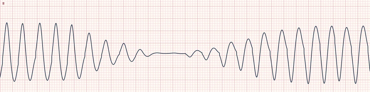
Example tracing, drawn by the lab's engine. Open the lab for a live, adjustable version.
At a glance
- Rate
- Usually 180–280/min
- Rhythm
- Irregular
- P waves
- Not visible
- QRS
- Wide, changing shape and axis beat to beat
Figures
Still images drawn by the lab's engine at 25 mm/s and 10 mm/mV, with the finding labeled. Select a figure to enlarge it.
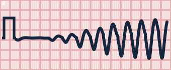
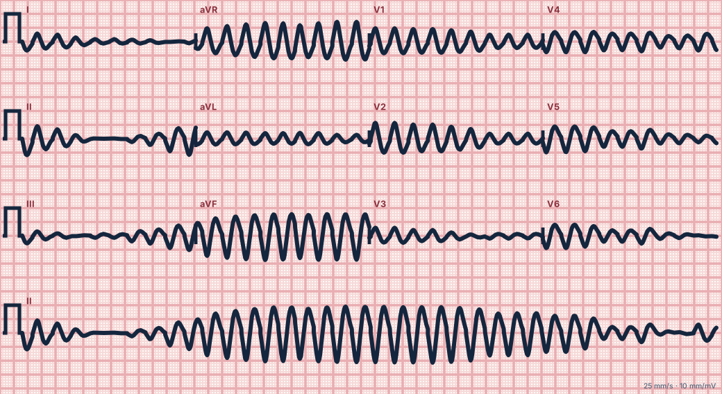
Scroll sideways to see the whole tracing, or select it to enlarge.
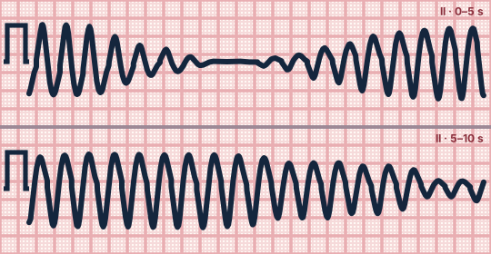
Scroll sideways to see the whole tracing, or select it to enlarge.
What the heart is doing
Several wavefronts or a shifting circuit activate the ventricles from continually changing directions, so each beat has a different shape. Output is poor and the rhythm tends to break down into fibrillation.
Why it looks this way
This is polymorphic VT because the fast, wide complexes vary continuously in shape and axis, without the smooth spindle pattern of torsades.
Why it matters
Highly unstable and quickly becomes VF. A sustained run needs an immediate unsynchronized shock, with or without a pulse, because the changing QRS prevents reliable synchronization. Then treat ischemia.
What to look for
- Wide complexes that keep changing height and direction, without a smooth repeating twist.
- The rhythm is fast and slightly irregular with no isoelectric baseline.
- Check the QT on any sinus beats beforehand: normal QT points to ischemia.
Criteria
- Rate
- Usually 180–280/min
- Rhythm
- Irregular
- P waves
- Not visible
- QRS
- Wide, changing shape and axis beat to beat
- Baseline QT
- Normal before the arrhythmia
Causes
- Acute myocardial ischemia or infarction
- Cardiomyopathy
- Brugada syndrome
- Catecholamine-triggered inherited VT
- Severe electrolyte disturbance
Can be mistaken for
- Torsades de pointes — smooth twisting amplitude with a long QT beforehand Compare
- Coarse ventricular fibrillation — no identifiable QRS complexes at all Compare
- Pre-excited atrial fibrillation — irregular but with a stable axis Compare
Advanced Down to the ion channel
What the cells are doing
Pacemaker, atrial and ventricular action potentials, in step with the tracing above, with sodium, calcium and potassium moving through their channels.
Frequently asked questions
What are the ECG criteria for polymorphic ventricular tachycardia?
Rate: usually 180–280/min. Rhythm: irregular. P waves: not visible. QRS: wide, changing shape and axis beat to beat. Baseline QT: normal before the arrhythmia.
How is polymorphic ventricular tachycardia identified on an ECG?
This is polymorphic VT because the fast, wide complexes vary continuously in shape and axis, without the smooth spindle pattern of torsades.
What can polymorphic ventricular tachycardia be mistaken for?
Torsades de pointes: smooth twisting amplitude with a long QT beforehand. Coarse ventricular fibrillation: no identifiable QRS complexes at all. Pre-excited atrial fibrillation: irregular but with a stable axis.
What causes polymorphic ventricular tachycardia?
Common causes: acute myocardial ischemia or infarction; cardiomyopathy; brugada syndrome; catecholamine-triggered inherited VT; and severe electrolyte disturbance.
Sources
This page does not list page-specific sources yet.
Every source the site cites, with links