
Ventricular
Draft — pending owner review
Bidirectional ventricular tachycardia
A rare VT in which the frontal QRS axis flips with every beat, so the limb-lead complexes alternate in direction. Think of digoxin toxicity or catecholaminergic polymorphic VT.
Last updated
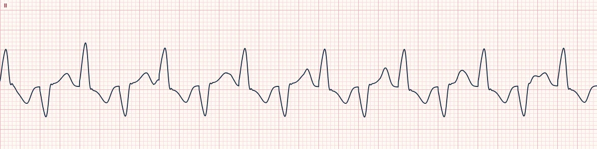
Example tracing, drawn by the lab's engine. Open the lab for a live, adjustable version.
At a glance
- Rhythm
- Regular wide-complex tachycardia
- Alternation
- Axis swings between left and right on alternate beats
- QRS
- Moderately wide, often RBBB-like in V1
- Pattern
- Strict beat-to-beat alternation, not a gradual twist
Figures
Still images drawn by the lab's engine at 25 mm/s and 10 mm/mV, with the finding labeled. Select a figure to enlarge it.
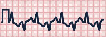
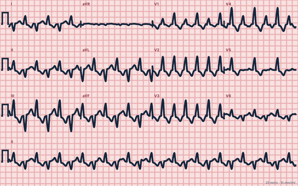
Scroll sideways to see the whole tracing, or select it to enlarge.
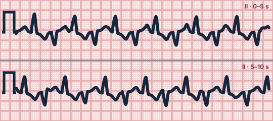
Scroll sideways to see the whole tracing, or select it to enlarge.
What the heart is doing
Calcium overload causes delayed afterdepolarizations that trigger beats from two sites, often near the left fascicles. They fire in turn, so the wavefront direction changes with every beat.
Why it looks this way
This is bidirectional VT because a regular wide tachycardia alternates its frontal axis with every beat, so the limb-lead complexes alternate in direction.
Why it matters
Strongly suggests digoxin toxicity or an inherited calcium-handling disorder, and can degenerate into polymorphic VT or VF. In digoxin toxicity check the potassium and seek antidote therapy; in CPVT adrenaline drives it.
What to look for
- In the limb leads one beat points up and the next points down, again and again.
- V1 often keeps the same RBBB-like shape on every beat.
- Ask about digoxin, plant toxins and events triggered by exercise or emotion.
Criteria
- Rhythm
- Regular wide-complex tachycardia
- Alternation
- Axis swings between left and right on alternate beats
- QRS
- Moderately wide, often RBBB-like in V1
- Pattern
- Strict beat-to-beat alternation, not a gradual twist
- Setting
- Digoxin toxicity, CPVT, aconite poisoning
Causes
- Digoxin toxicity
- Catecholaminergic polymorphic VT (CPVT)
- Aconite poisoning
- Andersen–Tawil syndrome
Can be mistaken for
- Torsades de pointes — the axis twists gradually over several beats Compare
- Electrical alternans — alternating height rather than direction, with a narrow QRS
- Ventricular bigeminy — every other beat is a normal sinus beat Compare
Advanced Down to the ion channel
What the cells are doing
Pacemaker, atrial and ventricular action potentials, in step with the tracing above, with sodium, calcium and potassium moving through their channels.
Frequently asked questions
What are the ECG criteria for bidirectional ventricular tachycardia?
Rhythm: regular wide-complex tachycardia. Alternation: axis swings between left and right on alternate beats. QRS: moderately wide, often RBBB-like in V1. Pattern: strict beat-to-beat alternation, not a gradual twist. Setting: digoxin toxicity, CPVT, aconite poisoning.
How is bidirectional ventricular tachycardia identified on an ECG?
This is bidirectional VT because a regular wide tachycardia alternates its frontal axis with every beat, so the limb-lead complexes alternate in direction.
What can bidirectional ventricular tachycardia be mistaken for?
Torsades de pointes: the axis twists gradually over several beats. Electrical alternans: alternating height rather than direction, with a narrow QRS. Ventricular bigeminy: every other beat is a normal sinus beat.
What causes bidirectional ventricular tachycardia?
Common causes: digoxin toxicity; catecholaminergic polymorphic VT (CPVT); aconite poisoning; and andersen–Tawil syndrome.
Sources
This page does not list page-specific sources yet.
Every source the site cites, with links