
Ventricular
Draft — pending owner review
Right ventricular outflow tract (RVOT) tachycardia
An idiopathic VT that starts in the right ventricular outflow tract of a structurally normal heart. The QRS has a left bundle branch block shape with an inferior axis.
Last updated
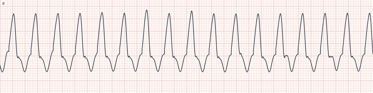
Example tracing, drawn by the lab's engine. Open the lab for a live, adjustable version.
At a glance
- QRS
- Wide, LBBB-like: dominant S wave in V1
- Axis
- Inferior: tall R waves in II, III and aVF
- Transition
- Late, usually V3–V4 or beyond
- Pattern
- Often repetitive bursts or frequent ectopic beats of the same shape
Figures
Still images drawn by the lab's engine at 25 mm/s and 10 mm/mV, with the finding labeled. Select a figure to enlarge it.
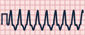
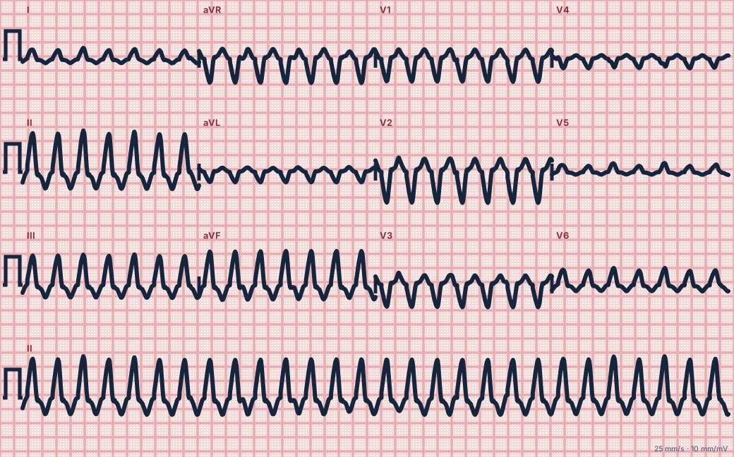
Scroll sideways to see the whole tracing, or select it to enlarge.
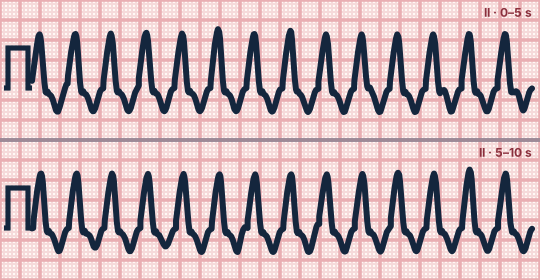
Scroll sideways to see the whole tracing, or select it to enlarge.
What the heart is doing
Adrenaline-driven, calcium-dependent triggered activity fires from the outflow tract high in the right ventricle. The wavefront runs down and to the left, so the inferior leads are positive and V1 is negative.
Why it looks this way
This is RVOT tachycardia because a wide LBBB-shaped QRS has an inferior axis, with tall R waves in II, III and aVF.
Why it matters
Usually benign and often curable by ablation, but the same shape occurs in arrhythmogenic right ventricular cardiomyopathy and in scar. It is called idiopathic only after structural disease is excluded; in the field it is managed like any VT.
What to look for
- A wide LBBB-shaped QRS with tall positive complexes in II, III and aVF.
- Runs that start and stop, often brought on by exercise or stress.
- Single ectopic beats between runs with exactly the VT's shape.
Criteria
- QRS
- Wide, LBBB-like: dominant S wave in V1
- Axis
- Inferior: tall R waves in II, III and aVF
- Transition
- Late, usually V3–V4 or beyond
- Pattern
- Often repetitive bursts or frequent ectopic beats of the same shape
- Heart
- Usually structurally normal
Causes
- Triggered activity in outflow-tract muscle
- Exercise or emotional stress
- Caffeine and stimulants
- Arrhythmogenic right ventricular cardiomyopathy (must be excluded)
Can be mistaken for
- Ventricular tachycardia, LBBB morphology — superior axis, typical of a lower right ventricular focus Compare
- SVT with LBBB — a typical LBBB shape with a brisk initial deflection
- Arrhythmogenic right ventricular cardiomyopathy — epsilon waves and T inversion in V1–V3 between runs Compare
Advanced Down to the ion channel
What the cells are doing
Pacemaker, atrial and ventricular action potentials, in step with the tracing above, with sodium, calcium and potassium moving through their channels.
Frequently asked questions
What are the ECG criteria for right ventricular outflow tract (RVOT) tachycardia?
QRS: wide, LBBB-like: dominant S wave in V1. Axis: inferior: tall R waves in II, III and aVF. Transition: late, usually V3–V4 or beyond. Pattern: often repetitive bursts or frequent ectopic beats of the same shape. Heart: usually structurally normal.
How is right ventricular outflow tract (RVOT) tachycardia identified on an ECG?
This is RVOT tachycardia because a wide LBBB-shaped QRS has an inferior axis, with tall R waves in II, III and aVF.
What can right ventricular outflow tract (RVOT) tachycardia be mistaken for?
Ventricular tachycardia, LBBB morphology: superior axis, typical of a lower right ventricular focus. SVT with LBBB: a typical LBBB shape with a brisk initial deflection. Arrhythmogenic right ventricular cardiomyopathy: epsilon waves and T inversion in V1–V3 between runs.
What causes right ventricular outflow tract (RVOT) tachycardia?
Common causes: triggered activity in outflow-tract muscle; exercise or emotional stress; caffeine and stimulants; and arrhythmogenic right ventricular cardiomyopathy (must be excluded).
Sources
This page does not list page-specific sources yet.
Every source the site cites, with links