
The waves
The U wave
A small wave after the T
A small positive wave after the T wave, best seen in V2 and V3 at slow rates. Prominent U waves are a classic sign of low potassium.
Last updated
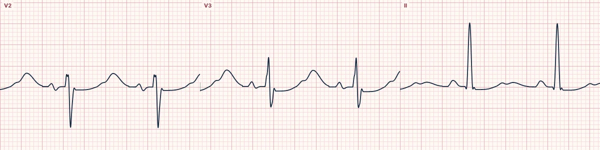
Example tracing, drawn by the lab's engine. Open the lab for a live, adjustable version.
At a glance
- Best seen
- V2–V3
- Normal size
- < 1–1.5 mm, about 10% of T
- Prominent
- Hypokalemia, bradycardia, drugs
Figures
Still images drawn by the lab's engine at 25 mm/s and 10 mm/mV, with the finding labeled. Select a figure to enlarge it.
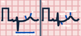
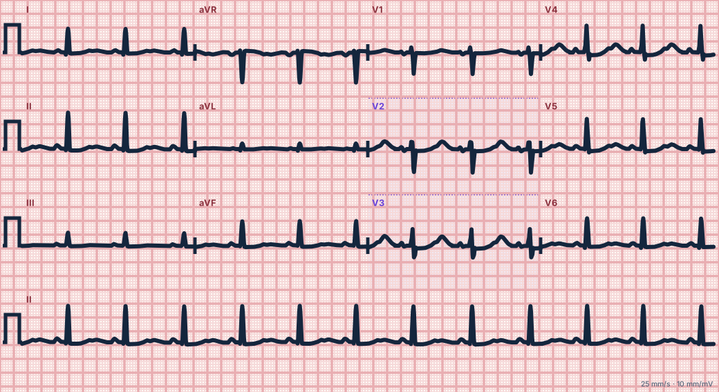
Scroll sideways to see the whole tracing, or select it to enlarge.
What it is
Its origin is still debated: delayed repolarization of the Purkinje fibres or mid-myocardial cells, or afterpotentials from the mechanical stretch of the ventricles. It normally points the same way as the T wave and is about a tenth of its height.
When it grows
Low potassium, bradycardia and some drugs make the U wave larger. In hypokalemia the T wave flattens and the U wave grows out of its tail, peaking about 0.1–0.15 s after it and often taller than the T in V2–V3, until the two merge, making the QT look long when it is really a QU interval.
Inverted U waves
A negative U wave in the chest leads is uncommon and points toward ischemia, hypertension or valve disease.
How to read it
- Look just after the T wave in V2 and V3.
- Do not include the U wave in the QT measurement.
Pitfalls
- A P wave of the next beat can look like a U wave at fast rates.
Key numbers
- Best seen
- V2–V3
- Normal size
- < 1–1.5 mm, about 10% of T
- Prominent
- Hypokalemia, bradycardia, drugs
Advanced Down to the ion channel
What the cells are doing
Pacemaker, atrial and ventricular action potentials, in step with the tracing above, with sodium, calcium and potassium moving through their channels.
Frequently asked questions
The U wave: what is the key idea?
A small positive wave after the T wave, best seen in V2 and V3 at slow rates. Prominent U waves are a classic sign of low potassium.
The U wave: what are the key numbers?
Best seen: V2–V3; Normal size: < 1–1.5 mm, about 10% of T; Prominent: Hypokalemia, bradycardia, drugs.
The U wave: how do you apply it to an ECG?
Look just after the T wave in V2 and V3. Do not include the U wave in the QT measurement.
The U wave: what are the common pitfalls?
A P wave of the next beat can look like a U wave at fast rates.
Sources
This page does not list page-specific sources yet.
Every source the site cites, with links