
Heart muscle and systemic disease
Draft — pending owner review
Hypothyroidism
Slow rates and small complexes
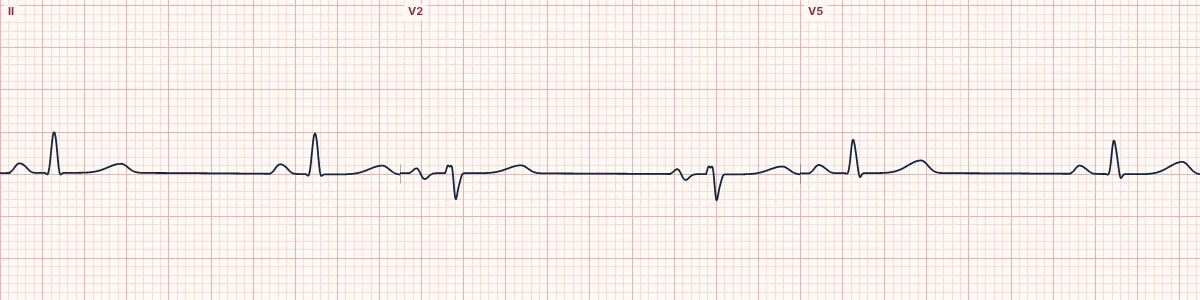
Low thyroid hormone slows the sinus node and can cause a pericardial effusion. Sinus bradycardia, low voltage, flattened or inverted T waves and a long QT are typical; myxedema coma adds severe bradycardia and hypothermia.
Common findings
Sinus bradycardia, low QRS voltage, flattened or inverted T waves, a prolonged QT and sometimes first-degree AV block or bundle branch block.
Myxedema coma
Marked bradycardia, low voltage, hypothermia with Osborn waves and QT prolongation that can lead to torsades de pointes.
Why it matters
Unexplained bradycardia, low voltage or a long QT deserve thyroid testing, especially in older people.
About the demonstration
The tracing shows sinus bradycardia with low QRS voltage; T-wave flattening and QT prolongation are not added.
How to read it
- Measure the rate, QRS voltage and QTc.
- Look for electrical alternans if an effusion is suspected.
- Look for Osborn waves when the temperature is low.
Pitfalls
- Low voltage is often put down to body habitus.
Frequently asked questions
Hypothyroidism: what is the key idea?
Low thyroid hormone slows the sinus node and can cause a pericardial effusion. Sinus bradycardia, low voltage, flattened or inverted T waves and a long QT are typical; myxedema coma adds severe bradycardia and hypothermia.
Hypothyroidism: what are the key numbers?
Rate: Sinus bradycardia; Voltage: Low (effusion, myxedema); QT: Prolonged; Severe: Hypothermia and Osborn waves in myxedema coma.
Hypothyroidism: how do you apply it to an ECG?
Measure the rate, QRS voltage and QTc. Look for electrical alternans if an effusion is suspected. Look for Osborn waves when the temperature is low.
Hypothyroidism: what are the common pitfalls?
Low voltage is often put down to body habitus.