
Ischemia & prior infarction
Draft — pending owner review
Left ventricular aneurysm (persistent ST elevation)
ST elevation that persists weeks after an anterior infarction, sitting over Q or QS waves, caused by a scarred, bulging segment of ventricle. It is easily mistaken for an acute STEMI.
Last updated
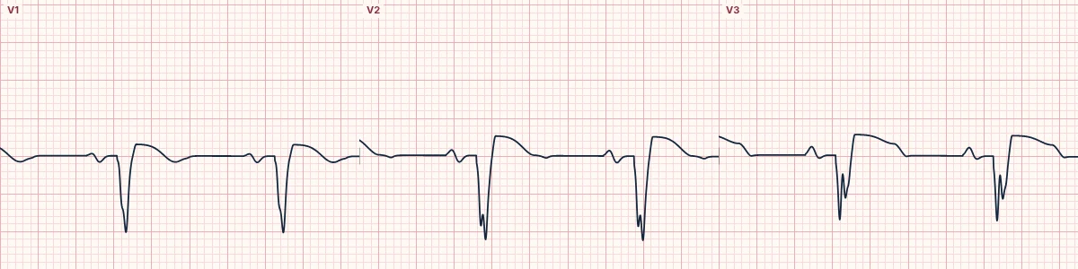
Example tracing, drawn by the lab's engine. Open the lab for a live, adjustable version.
At a glance
- QRS
- Q or QS waves in V1–V4
- ST segment
- Persistent elevation, usually convex, two weeks or more after MI
- T waves
- Small relative to the QRS
- T/QRS ratio
- Below 0.36 in every lead V1–V4 favors aneurysm; above it in any lead suggests acute MI
Figures
Still images drawn by the lab's engine at 25 mm/s and 10 mm/mV, with the finding labeled. Select a figure to enlarge it.
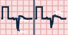
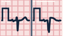
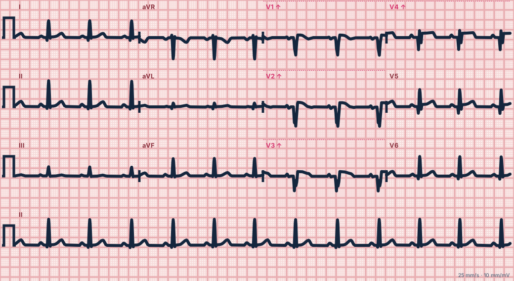
Scroll sideways to see the whole tracing, or select it to enlarge.
What the heart is doing
The thin, akinetic scar no longer depolarizes, leaving Q waves. Its abnormal wall tension and motion keep an injury-like current flowing, so the ST segment stays elevated long after the infarct.
Why it looks this way
This is a left ventricular aneurysm pattern because convex ST elevation sits over QS waves in V1–V3 while the T waves stay small relative to the QRS.
Why it matters
The danger is confusing it with a new STEMI, or the reverse; old ECGs and the T/QRS ratio help, and a new occlusion can still occur. Aneurysms predispose to heart failure, mural thrombus and ventricular tachycardia.
What to look for
- Find the Q or QS waves underneath the ST elevation.
- Divide T-wave height by QRS amplitude in V1–V4: small T waves point to an old aneurysm.
- Compare with old ECGs: unchanged ST elevation favors aneurysm.
Criteria
- QRS
- Q or QS waves in V1–V4
- ST segment
- Persistent elevation, usually convex, two weeks or more after MI
- T waves
- Small relative to the QRS
- T/QRS ratio
- Below 0.36 in every lead V1–V4 favors aneurysm; above it in any lead suggests acute MI
- Comparison
- Unchanged from earlier ECGs
Causes
- Prior large anterior infarction
- Late or no reperfusion of the infarct artery
- Prior inferior infarction (inferior aneurysm, less common)
Can be mistaken for
- Mid LAD occlusion — acute, with T waves large relative to the QRS Compare
- Prior anterior infarction — Q waves without persistent ST elevation Compare
- Benign early repolarization — no Q waves, and tall T waves Compare
Advanced Down to the ion channel
What the cells are doing
Pacemaker, atrial and ventricular action potentials, in step with the tracing above, with sodium, calcium and potassium moving through their channels.
Frequently asked questions
What are the ECG criteria for left ventricular aneurysm (persistent ST elevation)?
QRS: Q or QS waves in V1–V4. ST segment: persistent elevation, usually convex, two weeks or more after MI. T waves: small relative to the QRS. T/QRS ratio: below 0.36 in every lead V1–V4 favors aneurysm; above it in any lead suggests acute MI. Comparison: unchanged from earlier ECGs.
How is left ventricular aneurysm (persistent ST elevation) identified on an ECG?
This is a left ventricular aneurysm pattern because convex ST elevation sits over QS waves in V1–V3 while the T waves stay small relative to the QRS.
What can left ventricular aneurysm (persistent ST elevation) be mistaken for?
Mid LAD occlusion: acute, with T waves large relative to the QRS. Prior anterior infarction: Q waves without persistent ST elevation. Benign early repolarization: no Q waves, and tall T waves.
What causes left ventricular aneurysm (persistent ST elevation)?
Common causes: prior large anterior infarction; late or no reperfusion of the infarct artery; and prior inferior infarction (inferior aneurysm, less common).
Sources
This page does not list page-specific sources yet.
Every source the site cites, with links